45 simple microscope diagram with labels
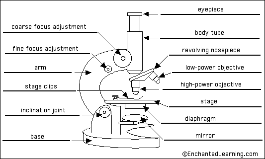
Parts of a Simple Microscope - Labeled (with diagrams) Parts of a Simple Microscope - Labeled (with diagrams) A simple microscope is a very first type of microscope ever created. It consists of simple parts and performs simple functions. Although there are now many advanced microscope types, some applications may still demand the use of a simple microscope. Labelled Diagram of Compound Microscope - Biology Discussion The below mentioned article provides a labelled diagram of compound microscope. Part # 1. The Stand: The stand is made up of a heavy foot which carries a curved inclinable limb or arm bearing the body tube. The foot is generally horse shoe-shaped structure (Fig. 2) which rests on table top or any other surface on which the microscope in kept.
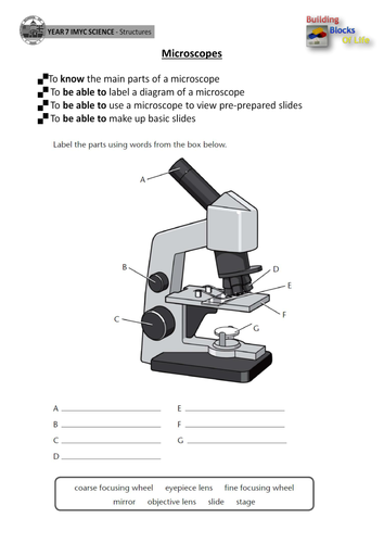
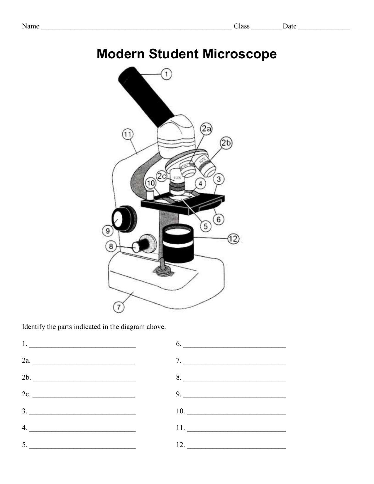
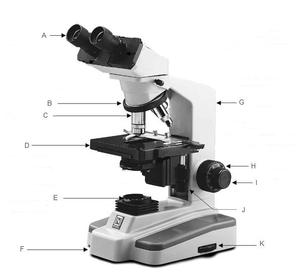
Microscope Labeling - The Biology Corner Students label the parts of the microscope in this photo of a basic laboratory light microscope. Can be used for practice or as a quiz. ... The type of microscope used in most science classes is the _____ microscope. 18. You should carry the microscope by the _____ and the _____. 19. The objectives are attached to what part of the microscope ...

Simple microscope diagram with labels
Microscope Types (with labeled diagrams) and Functions Simple microscope labeled diagram Simple microscope functions It is used in industrial applications like: Watchmakers to assemble watches Cloth industry to count the number of threads or fibers in a cloth Jewelers to examine the finer parts of jewelry Miniature artists to examine and build their work Also used to inspect finer details on products Anatomy Chart - How to Make Medical Drawings and Illustrations Pathologic anatomy focuses on how diseases affect and change the human body. Histology studies microscopic anatomy such as tissues and cells visible only under a microscope. Anatomy charts serve two main purposes: education in the form of anatomy worksheets and presentation in the form of simple healthcare illustrations. 16 Parts of a Compound Microscope: Diagrams and Video Once you have an understanding of the parts of the microscope it will be much easier to navigate around and begin observing your specimen, which is the fun part! The 16 core parts of a compound microscope are: Head (Body) Arm. Base. Eyepiece. Eyepiece tube.
Simple microscope diagram with labels. Electron microscope - Wikipedia An electron microscope is a microscope that uses a beam of accelerated electrons as a source of illumination. As the wavelength of an electron can be up to 100,000 times shorter than that of visible light photons , electron microscopes have a higher resolving power than light microscopes and can reveal the structure of smaller objects. Microscope Poster - Diagram with Labels | Teach Starter A poster containing a diagram with labels showing the key parts of a microscope. Use this educational classroom poster in your science lessons to highlight the key parts of a microscope. A lot of equipment is used in science experiments and it is important to know the names of and understand each part of the equipment and how it works. Microscope labeled diagram - slideshare.net Microscope labeled diagram Oct. 30, 2013 • 6 likes • 27,602 views Download Now Download to read offline Pisgah High School Follow 1. The Microscope Image courtesy of: Microscopehelp.com Basic rules to using the microscope 1. You should always carry a microscope with two hands, one on the arm and the other under the base. 2. Microscope Labeling - The Biology Corner 1) Start with scanning (the shortest objective) and only use the COARSE knob . Once it is focused… 2) Switch to low power (medium) and only use the COARSE knob . You may need to recenter your slide. Once it is focused.. 3) Switch to high power (long objective).
A Study of the Microscope and its Functions With a … Illuminator - Simple compound microscopes have a mirror that can be moved to adjust the amount of light that is focused on the specimen. However, some advanced types of compound microscopes have their own light source. ... These labeled microscope diagrams and the functions of its various parts, attempt to simplify the microscope for you ... Microscope Drawing Easy with Label - YouTube a great way to study is by using the blank version at the end of the video and practicing going over the parts of the microscope monocular vs binocular microscopes arm, stage, eye piece, body tube,... Microscope, Microscope Parts, Labeled Diagram, and … 19/01/2022 · Revolving Nosepiece or Turret: Turret is the part of the microscope that holds two or multiple objective lenses and helps to rotate objective lenses and also helps to easily change power. Objective Lenses: Three are 3 or 4 objective lenses on a microscope. The objective lenses almost always consist of 4x, 10x, 40x and 100x powers. The most common eyepiece lens is … Simple Microscope - Definition, Types, Working Principle … 1. Simple microscope comprises a biconvex lens used as a magnifying glass. Compound microscope comprises 2 or more convex lenses where one lens is the eyepiece and the other one is the objective lens. 2. Natural light is the source to see the object. An illuminator is a source to see the object. 3.
Label the Microscope Diagram | Download Scientific Diagram - ResearchGate the antibiogram of e. coli was investigated in different generations using eight antibiotic discs such as chloramphenicol (ch), streptomycin (s), gentamycin (g), ciprofloxacin (ci),... Simple Microscope Definition, Magnification, Parts And Uses - BYJUS To make a simple microscope with the help of water. Apparatus Required A glass of water Fuse wire Object to view (newspaper works well due to its fine print) Procedure Make a loop of the fuse wire around 2 mm wide. Dip it in water so that a drop is made in the loop. Hold it near to your eye and take a close look at the object you have chosen. Compound Microscope Parts, Functions, and Labeled Diagram Compound Microscope Parts, Functions, and Labeled Diagram Parts of a Compound Microscope Each part of the compound microscope serves its own unique function, with each being important to the function of the scope as a whole. Labeling the Parts of the Microscope | Microscope World Resources Labeling the Parts of the Microscope. This activity has been designed for use in homes and schools. Each microscope layout (both blank and the version with answers) are available as PDF downloads. You can view a more in-depth review of each part of the microscope here.
Biology Microscope Diagram Gcse - Micropedia
Microscope Parts, Function, & Labeled Diagram 24/12/2021 · Condenser. The condenser is to focus the light, which passes from the microscopic illuminator to the specimen. This condenser is located just below the diaphragm. This diaphragm is one of the important parts of the compound microscope which will help to get an accurate and sharp image. The condenser has a magnification power of 400X and above.
Microscope Labelled Diagram Gcse - Micropedia
Simple Microscope: Definition, Principle, Parts, And Uses A simple microscope is a rudimentary magnification device that is capable of visibly enlarging small objects, so they can be viewed and studied in better detail. It was invented in the late 16th century, and is still being widely used today. Simple microscopes have a wide range of applications in various fields.

Microscope-Diagram-Unlabeled.jpg (927×1200) | Science diagrams, Science printables, Biology labs
Free Microscope Worksheets for Simple Science Fun for Your Students 1. Parts of a Microscope . The first worksheet labels the different parts of a microscope, including the base, slide holder, and condenser. If you have a microscope, compare and contrast this worksheet to it.Also, your kids can color this microscope diagram in and read the words to each part of the microscope.
Permanent tissue: characteristics, types and functions - Online Biology Notes
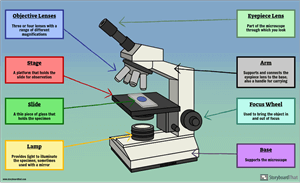
Compound Microscope Parts - Labeled Diagram and their Functions There are three major structural parts of a compound microscope. The head includes the upper part of the microscope, which houses the most critical optical components, and the eyepiece tube of the microscope. The base acts as the foundation of microscopes and houses the illuminator. The arm connects between the base and the head parts.

31 Label The Indicated Parts Of The Microscope - Labels For Your Ideas
Looking at the Structure of Cells in the Microscope ... Many light-microscope techniques are available for observing cells. Cells that have been fixed and stained can be studied in a conventional light microscope, while antibodies coupled to fluorescent dyes can be used to locate specific molecules in cells in a fluorescence microscope. Living cells can be seen with phase-contrast, differential ...

Simple Microscope Diagram Blank - Micropedia
Label the microscope — Science Learning Hub All microscopes share features in common. In this interactive, you can label the different parts of a microscope. Use this with the Microscope parts activity to help students identify and label the main parts of a microscope and then describe their functions. Drag and drop the text labels onto the microscope diagram.

Microscope Labelled Diagram Ks3 - Micropedia
Microscope With Labels clip art | Microscope parts, Scientific method ... Microscope With Labels clip art | Microscope parts, Scientific method, Science diagrams From clker.com vector clip art online, royalty free & public domain Download Clker's Microscope With Labels clip art and related images now. Multiple sizes and related images are all free on Clker.com. D Dixie Tsutsaeva 2k followers More information

Microscope With Labels Clip Art at Clker.com - vector clip art online, royalty free & public domain
Fluorescence Resonance Energy Transfer (FRET) Microscopy Presented in Figure 3 is a Jablonski diagram illustrating the coupled transitions involved between the donor emission and acceptor absorbance in fluorescence resonance energy transfer. Absorption and emission transitions are represented by straight vertical arrows (green and red, respectively), while vibrational relaxation is indicated by wavy ...

Histology: Epithelial Tissue: Simple Cuboidal | Human anatomy and physiology, Anatomy and ...
Microscope Poster - Diagram with Labels | Teach Starter A poster containing a diagram with labels showing the key parts of a microscope. In Science it is important that students know how to use a variety of tools when conducting scientific experiments and inquiry. This poster focuses on the microscope and highlights its key parts. There are two print options available for this poster:
Print Anatomy Midterm 2 flashcards | Easy Notecards
A physical wiring diagram for the human immune system | Nature Aug 03, 2022 · For imaging, a PerkinElmer Opera Phenix automated spinning-disk confocal microscope was used and each well of a 348-well plate was imaged at 20× magnification with 5 × 5 non-overlapping images ...

Microscope With Labels Clip Art at Clker.com - vector clip art online, royalty free & public domain
Parts of a microscope with functions and labeled … Structural parts of a microscope and their functions Figure created with biorender.com Figure: Diagram of parts of a microscope There are three structural parts of the microscope i.e. head, base, and arm. Head - This is also known as the body. It carries the optical parts in the upper part of the microscope. Base - It acts as microscopes support.

#39 Structure of transport tissues in plants | Biology Notes for A level
Draw a neat labeled diagram for the formation of an image in a simple ... A simple microscope is a convex lens where in the image is kept between the focus of the lens so that the image formed is virtual and magnified. While drawing make sure the symmetry is taken into consideration. Complete step by step answer: Here is the labeled diagram outlining a simple microscope. To draw the ray diagram, place the object ...
38 Microscope Diagram To Label - Labels 2021
Fluorescence - Wikipedia Fluorescence is the emission of light by a substance that has absorbed light or other electromagnetic radiation.It is a form of luminescence.In most cases, the emitted light has a longer wavelength, and therefore a lower photon energy, than the absorbed radiation.
Parts of the Microscope Flashcards | Easy Notecards
Simple Squamous Epithelium under a Microscope with a Labeled Diagram ... Simple columnar epithelium labeled. This is a labeled diagram of a simple columnar epithelium under a light microscope. I tried to show you both ciliated and nonciliated simple columnar epithelium. These diagrams show the cilia on the cell surface, rectangular cell, and elongated nucleus.
Microscope Diagram Labelled - Micropedia
Simple Microscope – Parts, Functions, Diagram and Labelling Simple Microscope - Parts, Functions, Diagram and Labelling By Editorial Team March 7, 2022 A microscope is one of the commonly used equipment in a laboratory setting. A microscope is an optical instrument used to magnify an image of a tiny object; objects that are not visible to the human eyes. Table of Contents

A Study of the Microscope and its Functions With a Labeled Diagram | Students, Homeschool and ...
Simple Microscope - Diagram (Parts labelled), Principle, Formula and Uses Parts of a Simple Microscope A simple microscope consists of Optical parts Mechanical parts Labeled Diagram of simple microscope parts Optical parts The optical parts of a simple microscope include Lens Mirror Eyepiece Lens A simple microscope uses biconvex lens to magnify the image of a specimen under focus.
Post a Comment for "45 simple microscope diagram with labels"