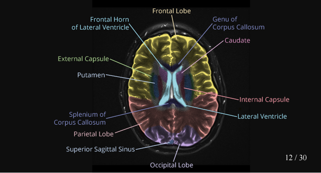
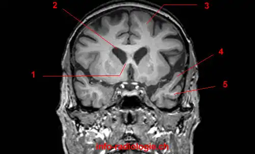
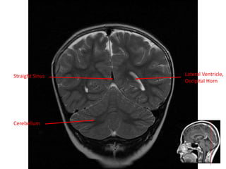
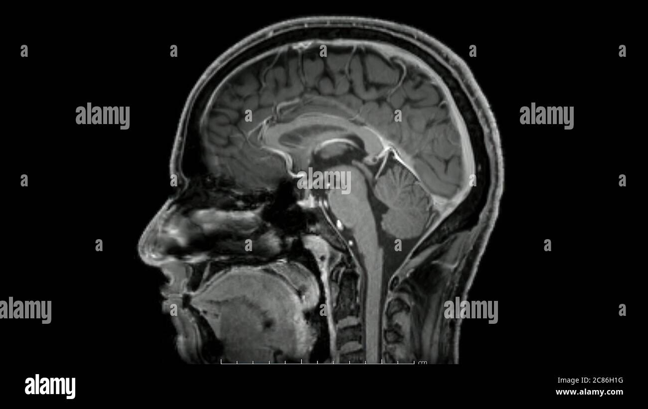
42 brain mri with labels
CPT Code for MRI Brain, Breast, Lumbar Spine and Shoulder Find below the latest Radiology CPT codes for for MRI of Brain, Breast, Lumbar Spine and Shoulder: CPT Codes for MRI Lumbar spine In human Lumbar spine is represented by the 5 vertebrae in between the ribcage and the pelvis forming the largest segment of the vertebral column. Depending on the condition that one is treated on these parts of the ... Labels · Th3NiKo/Brain-MRI-segmentation-unet · GitHub Training U-net segmentation cnn model for brain anomalous parts detection. - Labels · Th3NiKo/Brain-MRI-segmentation-unet
UCLA Brain Mapping Center - ICBM Template To view both the structural MRI and the labels launch the program typing Display icbm_template.mnc -label icbm_labels_corrected.mnc. The opacity of the labels can be set in the Colour Coding menu. The number of each label appears at the bottom left of the orthogonal views window.
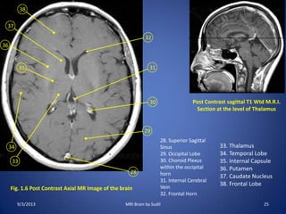
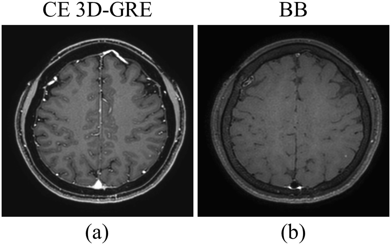
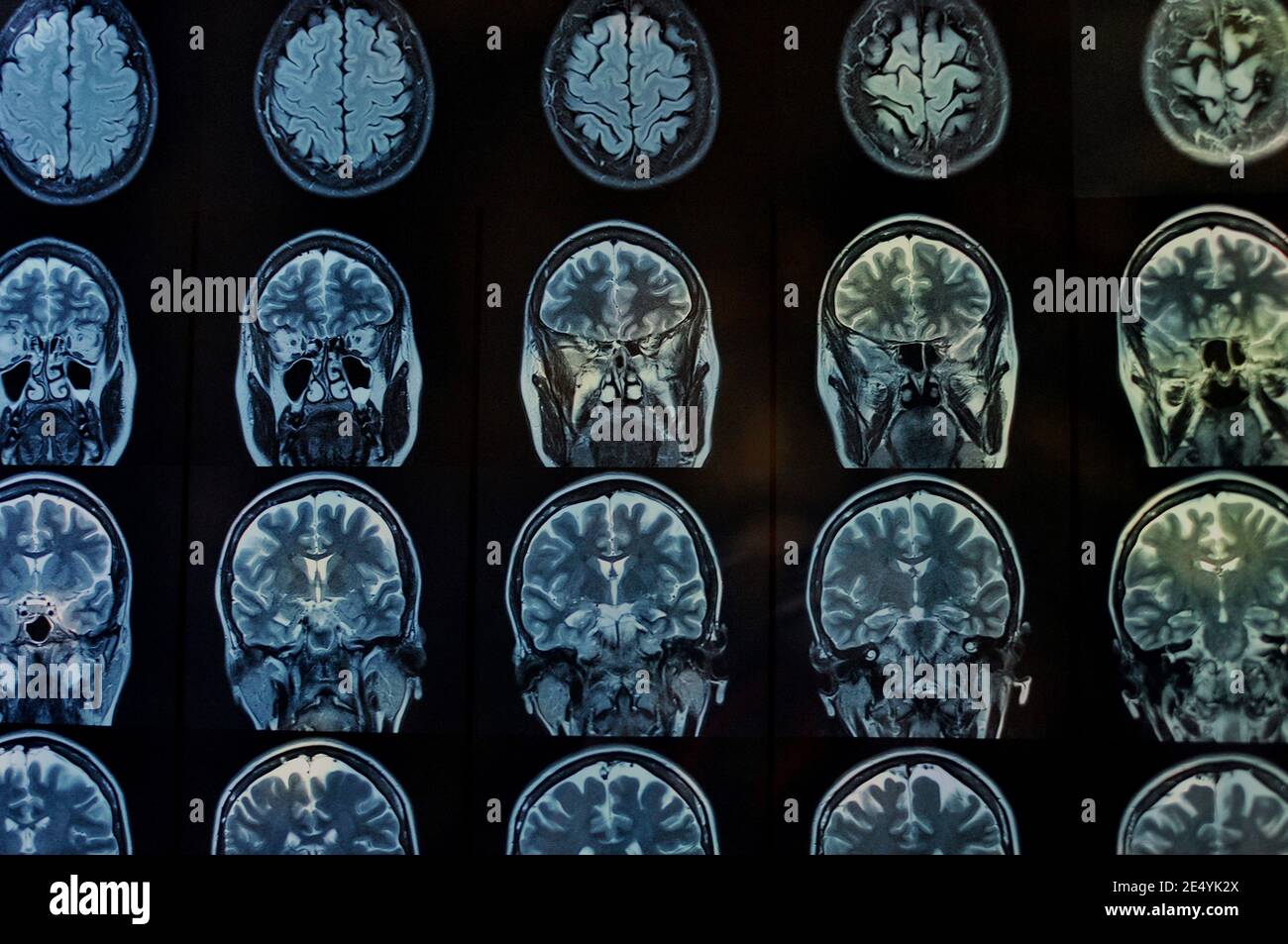
Brain mri with labels
NITRC: Manually Labeled MRI Brain Scan Database: Tool/Resource Info Manually Labeled MRI Brain Scan Database Visit Website Image 1 of 3 Click for more. This is a continuously growing and improving database of high-quality neuroanatomically labeled MRI brain scans, created not by an algorithm, but by neuroanatomical experts. All results are checked and corrected. Deep learning to automate the labelling of head MRI datasets for ... manually labelling mri scans appears to be particularly laborious due to (1) the superior soft-tissue contrast of mri which enables more refined diagnoses compared with other imaging modalities such as computed tomography; and (2) the use of multiple, complementary imaging sequences so that a larger number of images must be scrutinised per … Magnetic Resonance Imaging (MRI) of the Spine and Brain Magnetic resonance imaging (MRI) is a diagnostic procedure that uses a combination of a large magnet, radiofrequencies, and a computer to produce detailed images of organs and structures within the body. Unlike X-rays or computed tomography (CT scans), MRI does not use ionizing radiation. Some MRI machines look like narrow tunnels, while others ...
Brain mri with labels. RSNA-ASNR-MICCAI Brain Tumor Segmentation (BraTS) Challenge ... Participants are free to choose whether they want to focus only on one or both tasks. Task 1: Brain Tumor Segmentation in mpMRI scans. The participants are called to address this task by using the provided clinically-acquired training data to develop their method and produce segmentation labels of the different glioma sub-regions. brain anatomy | MRI coronal brain anatomy | free MRI cross sectional ... ELBOW AXIAL. WRIST AXIAL. WRIST CORONAL. KNEE CORONAL. KNEE SAGITTAL. ARTERIES UPPER LEG. ARTERIES LOWER LEG. This MRI brain coronal cross sectional anatomy tool is absolutely free to use. Use the mouse scroll wheel to move the images up and down alternatively use the tiny arrows (>>) on both side of the image to move the images. Researchers automate brain MRI image labeling, more than ... - ScienceDaily Researchers have automated brain MRI image labeling, needed to teach machine learning image recognition models, by deriving important labels from radiology reports and accurately assigning them to... Frontiers | 101 Labeled Brain Images and a Consistent Human Cortical ... Labeled anatomical subdivisions of the brain enable one to quantify and report brain imaging data within brain regions, which is routinely done for functional, diffusion, and structural magnetic resonance images (f/d/MRI) and positron emission tomography data.
Spinal cord injury - Wikipedia CT gives greater detail than X-rays, but exposes the patient to more radiation, and it still does not give images of the spinal cord or ligaments; MRI shows body structures in the greatest detail. Thus it is the standard for anyone who has neurological deficits found in SCI or is thought to have an unstable spinal column injury. CaseStacks.com - MRI Brain Anatomy Labeled scrollable brain MRI covering anatomy with a level of detail appropriate for medical students. Show/Hide Labels. MRI Brain Anatomy. Back to Anatomy Overview. ... Labelled radiographs and CT/MRI series teaching anatomy with a level of detail appropriate for medical students and junior residents. Pelvis. Pelvic MRI anatomy Labeled imaging anatomy cases | Radiology Reference Article ... This article lists a series of labeled imaging anatomy cases by body region and modality. Brain CT head: non-contrast axial CT head: non-contrast coronal CT head: non-contrast sagittal CT head: angiogram axial CT head: angiogram coronal CT... Whole Brain Perfusion Measurements Using Arterial Spin ... The MB ASL technique is an effective method to evaluate whole brain perfusion because it minimizes the temporal spread of labeled spins across slices, resulting in more accurate perfusion measurements.
Brain lobes - annotated MRI | Radiology Case | Radiopaedia.org Introduction to CT Brain by Dr Sally Ayesa Head (brain-eye-ear-nose-mouth) by Razan Ali Rawashdeh; 2021 by khaledawad; Anatomy by Dr Marwan El Toukhy; teaching cases by Mohamed Emad Shehata; Forensic cases by Dr Thomas M. Anderson Neuroanatomy Review by Dr. Francisco Gallegos; AAA-AnatomyBible by Dr Ian Bickle Brain by Nurudeen Olugbemiga Jimoh 2,831 Labeled brain anatomy Images, Stock Photos & Vectors - Shutterstock Find Labeled brain anatomy stock images in HD and millions of other royalty-free stock photos, illustrations and vectors in the Shutterstock collection. Thousands of new, high-quality pictures added every day. Automated MRI image labelling processes 100,000 brain exams in under 30 ... Researchers from the School of Biomedical Engineering & Imaging Sciences at King's College London have automated brain MRI image labeling, needed to teach machine learning image recognition models,... Brain Cell Data Center (BCDC) - Brain Cell Data Center (BCDC) Brain tissues are imaged and spatially registered using MRI, and sampling regions (visual cortex or BA17) are processed for single cell assays. Gene expression and chromatin accessible site mapping permits unbiased clustering of nuclei (visualized using t-SNE) and cell type annotation.
MRI Brain Atlas - University of Minnesota This web app Atlas is intended for veterinary students and radiologists seeking quick access to canine brain anatomy through a mobile device. Via a toggle button, either MRI images or approximately comparable Brain Transection images may be viewed with or without labels. Navigation & Labels.
101 Labeled Brain Images and a Consistent Human Cortical Labeling ... Labeled anatomical subdivisions of the brain enable one to quantify and report brain imaging data within brain regions, which is routinely done for functional, diffusion, and structural magnetic resonance images (f/d/MRI) and positron emission tomography data.
What Does a Brain MRI Show? • San Diego Health What does a brain MRI show? The answer is, unfortunately, not very. MRI scans (magnetic resonance imaging) have been around for decades, and the technology has been steadily improving. Today, a brain MRI test can identify whether or not a person has a stroke, or if the person has suffered a traumatic brain injury, or if the person is suffering ...
Deep learning from MRI-derived labels enables automatic brain tissue ... Deep learning from MRI-derived labels enables automatic brain tissue classification on human brain CT Neuroimage. 2021 Dec 1;244:118606. doi: 10.1016/j ... Our proposed model predicted brain tissue classes accurately from unseen CT images (Dice coefficients of 0.79, 0.82, 0.75, 0.93 and 0.98 for GM, WM, CSF, brain volume and ICV, respectively). ...
MRI anatomy | free MRI axial brain anatomy - Mrimaster.com This MRI brain cross sectional anatomy tool is absolutely free to use. Use the mouse scroll wheel to move the images up and down alternatively use the tiny arrows (>>) on both side of the image to move the images.
Atlas of BRAIN MRI - W-Radiology Brain magnetic resonance imaging (MRI) is a common medical imaging method that allows clinicians to examine the brain's anatomy (1). It uses a magnetic field and radio waves to produce detailed images of the brain and the brainstem to detect various conditions (2).
Brain: Atlas of human anatomy with MRI - e-Anatomy - IMAIOS MRI Atlas of the Brain. This page presents a comprehensive series of labeled axial, sagittal and coronal images from a normal human brain magnetic resonance imaging exam. This MRI brain cross-sectional anatomy tool serves as a reference atlas to guide radiologists and researchers in the accurate identification of the brain structures.
Brain MRI Images for Brain Tumor Detection | Kaggle Brain MRI Images for Brain Tumor Detection. Brain MRI Images for Brain Tumor Detection. Data. Code (246) Discussion (8) Metadata. About Dataset. No description available. Health Biology Classification Computer Vision Deep Learning. Edit Tags. close. search. Apply up to 5 tags to help Kaggle users find your dataset.
Labeled MRI Brain Scans - Neuromorphometrics We can also label scans that you provide and we are very interested in labeling white matter anatomy as seen in diffusion-weighted MRI scans. If you want an aggregate version of our data, we can provide it as a probabilistic atlas. The cost to label a single scan is $2449 (USD).
Mail Online Videos: Top News & Viral Videos, Clips & Footage ... Oct 05, 2022 · Check out the latest breaking news videos and viral videos covering showbiz, sport, fashion, technology, and more from the Daily Mail and Mail on Sunday.
Brain MRI Segmentation Using FCM (Labeling) - Stack Overflow This can be accomplished with label2rgb as the documentation suggests here. I would probably use this form: RGB = label2rgb (L, map) Where map is a colormap. If you pass the same map to each slice's call to label2rgb the labels will be returned with the same colors. This can also be implemented relatively easily.
Brain MRI Segmentation Using Pretrained 3-D U-Net Network Download Brain MRI and Label Data This example uses a subset of the CANDI data set [ 2] [ 3 ]. The subset consists of a brain MRI volume and the corresponding ground truth label volume for one patient. Both files are in the NIfTI file format. The total size of the data files is ~32 MB. Create a folder in which to store the data set.
Head MRI: Purpose, Preparation, and Procedure - Healthline A head MRI is a useful tool for detecting a number of brain conditions, including: aneurysms, or bulging in the blood vessels of the brain; multiple sclerosis; spinal cord injuries; hydrocephalus ...
Brain MRI segmentation | Kaggle This dataset contains brain MR images together with manual FLAIR abnormality segmentation masks. The images were obtained from The Cancer Imaging Archive (TCIA). They correspond to 110 patients included in The Cancer Genome Atlas (TCGA) lower-grade glioma collection with at least fluid-attenuated inversion recovery (FLAIR) sequence and genomic ...
Brain MRI: How to read MRI brain scan | Kenhub MRI is the most sensitive imaging method when it comes to examining the structure of the brain and spinal cord. It works by exciting the tissue hydrogen protons, which in turn emit electromagnetic signals back to the MRI machine. The MRI machine detects their intensity and translates it into a gray-scale MRI image.
101 labeled brain images and a consistent human cortical ... - PubMed given how difficult it is to label brains, the mindboggle-101 dataset is intended to serve as brain atlases for use in labeling other brains, as a normative dataset to establish morphometric variation in a healthy population for comparison against clinical populations, and contribute to the development, training, testing, and evaluation of …
Cross-sectional anatomy of the brain - e-Anatomy - IMAIOS An MRI was performed on a healthy subject, with several acquisitions with different weightings: spin-echo T1, T2 and FLAIR, T2 gradient-echo, diffusion, and T1 after gadolinium injection. We obtained 24 axial slices of the normal brain.
Magnetic Resonance Imaging (MRI) of the Spine and Brain Magnetic resonance imaging (MRI) is a diagnostic procedure that uses a combination of a large magnet, radiofrequencies, and a computer to produce detailed images of organs and structures within the body. Unlike X-rays or computed tomography (CT scans), MRI does not use ionizing radiation. Some MRI machines look like narrow tunnels, while others ...
Deep learning to automate the labelling of head MRI datasets for ... manually labelling mri scans appears to be particularly laborious due to (1) the superior soft-tissue contrast of mri which enables more refined diagnoses compared with other imaging modalities such as computed tomography; and (2) the use of multiple, complementary imaging sequences so that a larger number of images must be scrutinised per …
NITRC: Manually Labeled MRI Brain Scan Database: Tool/Resource Info Manually Labeled MRI Brain Scan Database Visit Website Image 1 of 3 Click for more. This is a continuously growing and improving database of high-quality neuroanatomically labeled MRI brain scans, created not by an algorithm, but by neuroanatomical experts. All results are checked and corrected.





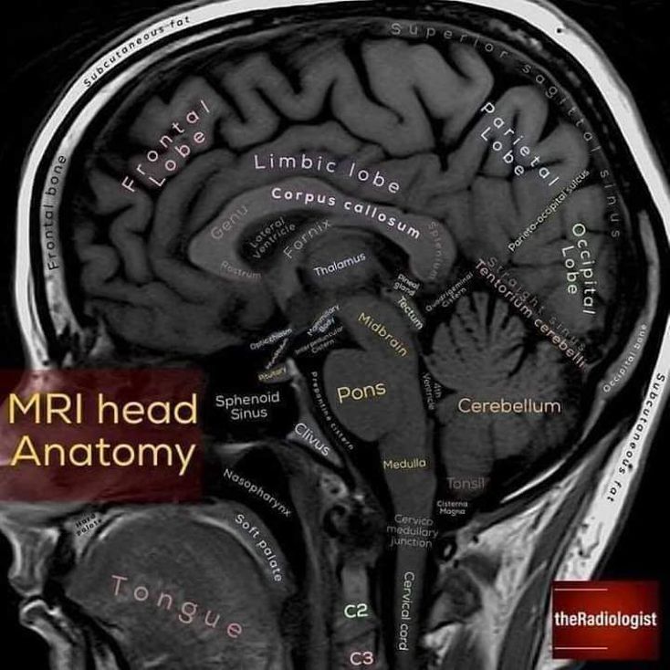


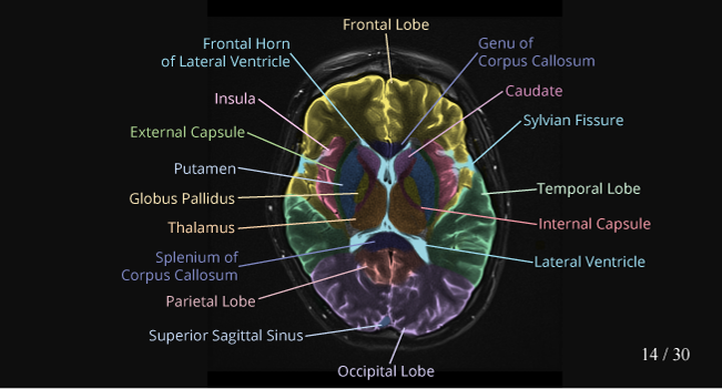
:watermark(/images/watermark_only_sm.png,0,0,0):watermark(/images/logo_url_sm.png,-10,-10,0):format(jpeg)/images/anatomy_term/thalamus-12/JikayR0budNwnh0ynTMMA_Thalamus.jpg)
:background_color(FFFFFF):format(jpeg)/images/library/13518/xLkcLnsMMPx5TgdQElAqvg_mri-t2-axial-thalamus-level_latin.jpg)

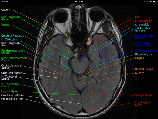

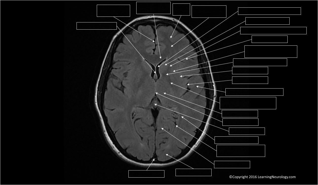






Post a Comment for "42 brain mri with labels"